Day 2 :
Keynote Forum
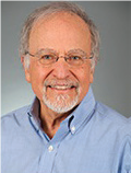
Larry I. Benowitz,
Harvard Medical School, USA.
Keynote: Optic nerve regeneration: regulation by amacrine cells, nitric oxide, and zinc
Time : 09:30-10:10

Biography:
Larry Benowitz is a Professor of Surgery and Ophthalmology. His research interests involve understanding the mechanisms that underlie cell death and regenerative failure after CNS injury, and developing methods to preserve damaged neurons and promote the rewiring of neural circuitry.
Abstract:
Retinal Ganglion Cells (RGCs), the neurons that project visual information from the eye to the brain, cannot regenerate their axons once the optic nerve has been injured and soon begin to die. This failure has dire consequences for victims of traumatic or ischemic nerve damage or degenerative diseases such as glaucoma. Our lab and others have recently identified methods that enable some RGCs to regenerate axons from the eye to the brain, yet most RGCs go on to die and only a small fraction of surviving RGCs regenerate their axons. These findings imply the existence of other major suppressors of RGC survival and axon regeneration. We recently identified mobile zinc (Zn2+) one such factor. Within an hour after optic
nerve injury, Zn2+ increases dramatically in synaptic vesicles of amacrine cells (ACs), the inhibitory interneurons of the retina, then transfers slowly to injured RGCs. Zn2+ chelation leads to the persistent survival of many RGCs and to appreciable axon regeneration, with a therapeutic window of several days. New results show that Zn2+ elevation is induced by nitric oxide (NO), a gaseous signal that is generated in a small population of ACs via glutamate-dependent activation of the enzyme NO synthase-1 (NOS1). A novel fl uorescent NO sensor reveals that retinal NO levels increase within 30 minutes of optic nerve damage. NO or a derivative thereof probably liberates Zn2+ from proteins such as metallothioneins via nitrosylation of Zn2+- binding cysteine residues. Surprisingly, we also find that NO has a second, positive effect on optic nerve regeneration through a cGMP-dependent pathway. Besides eliminating Zn2+ accumulation in the retina, AC-specific deletion of NOS1 blocked the regeneration that would otherwise have occurred upon Zn2+ chelation. Conversely, elevation of NO with the NO donor DETANONOate or prevention of cGMP degradation was sufficient to induce axon regeneration. Thus, NO generated by NOS1 in a small population of ACs is responsible for the deleterious elevation of Zn2+ after optic nerve regeneration, but also exerts a positive effect on optic nerve regeneration via cGMP signaling.
Keynote Forum
Chih-Yu Chen
Keynote: Augmented superior rectus transposition with intraoperative botulinum toxin injection for large angle chronic six nerve palsy: A case report

Biography:
Chih-Yu Chen has completed his PhD at the age of 25 years from Taipei Medical University and had obtained his Master degree of Medical Science from National Taiwan University later. He is currently working in the field of Pediatric Ophthalmology and Strabismus at Taipei Municipal Wanfang Hospital (Managed by Taipei
Medical University), Taiwan.
Abstract:
A case of 54 years-old female patient, presented with 75 ΔPD esotropia, combined with obvious hypotropia of right eye in primary position. She was a case of right traumatic six nerve palsy for three years with chin-up and face turn to right. There was a filtration bleb on the upper limbus of right eye due to acute attack of PACG after the trauma. Augmented superior rectus transposition with intraoperative botulinum toxin injection of right medial rectus, combined with bilateral medial rectus muscle recessions. (od 6.5 mm, os 4.5 mm) on December 2017. Complete ptosis with lateral gaze limitation (-2) (od) and nasal limitation (-1) (ou) was found two weeks aft er the operation. Later, 18-20 PD exotropia (od) was found with slight hypertropia and ptosis (3+) aft er one month. Two months later, there was no exotropia or esotropia, just six prism-diopters hypertropia and slight degree of ptosis was found. Four months later, orthotropia in primary position and no more ptosis. A gain of 15 degree lateral gaze across the midline was achieved after the SRT surgery compared complete paralysis before the operation. There was a final exoshift of 75 prism-diopters and a successful outcome was achieved in this case. Augmented Superior Rectus Transposition (SRT) with intraoperative botulinum toxin injection maybe a selective method for the treatment of large angle chronic six nerve palsy.
